
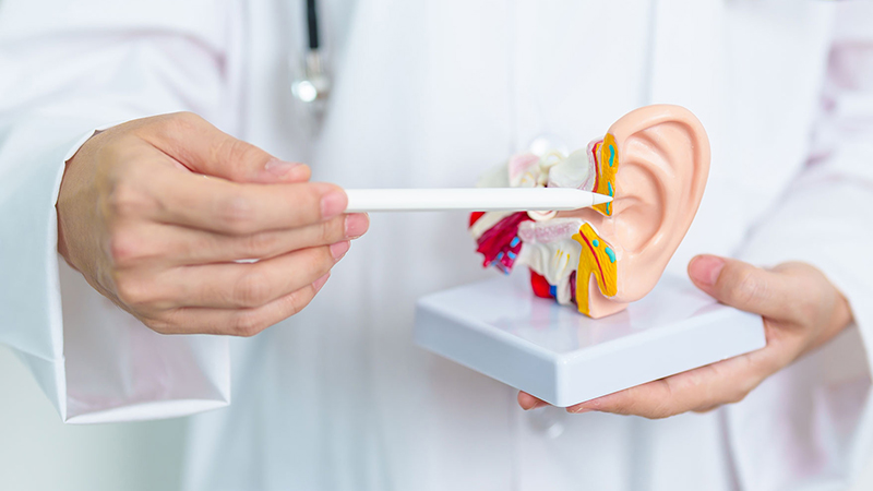
Otosclerosis is a cause of slowly progressive hearing loss in which abnormal bone growth restricts the movement of the tiny hearing bones in the middle ear. It is sometimes described as “hardening” or “calcification” of the middle ear, but it is not actually a buildup of calcium; it is a disorder of the way bone is remodeled. With a correct diagnosis, hearing can be improved significantly in most patients with options such as hearing aids or stapedotomy surgery.
What Is Otosclerosis?
Sound makes the eardrum vibrate, and three tiny middle ear bones, the malleus (hammer), incus (anvil) and stapes (stirrup), pass these vibrations to the inner ear. The stapes, the last link, sits with its base (footplate) in the oval window, the entrance to the inner ear, and moves like a piston to transmit sound into the inner ear fluids.
In otosclerosis, new bone tissue, spongy at first and then hardening, forms in the bony capsule surrounding the inner ear, especially around the oval window. This tissue fixes the stapes footplate in place and prevents vibrations from reaching the inner ear. The result is conductive hearing loss. If the disease also affects the inner ear itself, sensorineural (nerve-type) loss is added; when both are present, it is called mixed hearing loss.
Symptoms usually appear between the ages of 15 and 45, and the condition is about twice as common in women as in men. Most patients eventually have both ears affected, although the two ears may be involved to different degrees and at different times. It is more common in people of white European descent.
Otosclerotic bone changes do not always cause hearing loss and can be found in people with normal hearing; symptoms appear only when the oval window and stapes are involved. In cochlear otosclerosis, the form that affects the inner ear capsule, sensorineural hearing loss is more prominent and the course can be more complex.
Causes of Otosclerosis
The exact cause of otosclerosis is not known. Current evidence suggests that it arises from a combination of environmental and hormonal factors on a background of genetic susceptibility. The main risk factors are:
- Family history: Many patients have relatives with similar hearing loss; studies report a family history in roughly a third to two-thirds of cases. The condition can sometimes skip generations.
- Female sex and hormones: Hearing loss often begins or speeds up during pregnancy, which suggests that hormones play a role.
- Viral factors: Some research has suggested that the measles virus may trigger the bone changes. Some populations have reported fewer cases since vaccination became widespread.
- Age: Symptoms usually start in young adulthood and progress over the years.
Otosclerosis is not contagious, and it is not directly related to noise, ear infections or ear cleaning habits.
Symptoms of Otosclerosis
The hallmark is hearing loss that creeps up over months and years. With no pain or discharge, people often notice it only when the loss becomes obvious. Common symptoms include:
- Gradually increasing hearing loss in one or both ears, with whispers and low-pitched sounds especially hard to hear
- Tinnitus (ringing, humming or buzzing in the ear)
- Understanding speech better in noisy surroundings than in quiet ones (paracusis of Willis)
- Hearing one’s own voice loudly from the inside, and therefore tending to speak softly
- Difficulty hearing surrounding sounds while chewing
- Less often, mild unsteadiness or dizziness
When the inner ear is also affected, hearing loss deepens, understanding speech becomes harder and tinnitus may become more noticeable. This decline in hearing can affect social relationships, work life and self-confidence.
How Is Otosclerosis Diagnosed?
In otosclerosis, the eardrum usually looks completely normal, so diagnosis relies on interpreting the examination together with hearing tests. During the active phase of the disease, a reddish blush behind the eardrum, known as Schwartze’s sign, can rarely be seen.
The assessments used in diagnosis include:
- Tuning fork tests: Simple tests with vibrating metal forks show that the hearing loss is conductive and which ear is more affected.
- Audiometry: Air and bone conduction hearing thresholds are measured. The gap between them indicates conductive loss, and a typical dip in bone conduction around 2000 Hz (Carhart notch) supports the diagnosis.
- Tympanometry and acoustic reflex testing: Eardrum mobility is usually normal or slightly reduced. Because the stapes is fixed, the acoustic reflex usually cannot be recorded.
- CT scan: High-resolution CT of the temporal bone can show changes around the oval window and in the inner ear capsule, and it also helps with surgical planning.
These tests distinguish otosclerosis from look-alike conditions such as middle ear fluid, a break in the hearing bone chain or congenital fixation of the hearing bones.
When surgery is considered, hearing levels in both ears, speech discrimination and inner ear reserve are assessed in detail, along with general health, medications and previous ear surgery. This shows which ear to operate on first and what hearing gain is realistic. Patients traveling from abroad are encouraged to bring previous audiograms, CT images and reports, and a list of their current medications.
Otosclerosis Treatment
Treatment depends on the degree of hearing loss, whether one or both ears are affected, inner ear status, age and expectations. For mild loss, monitoring with regular hearing tests may be enough.
Medication
No medication reverses otosclerosis. In some patients whose inner ear is affected and whose hearing is declining quickly, the doctor may consider drugs such as sodium fluoride to slow bone remodeling. These do not restore lost hearing, and their effect on progression is limited.
Hearing Aids
A hearing aid is an effective and safe option for patients who do not want surgery, whose health does not allow it or who hear with only one ear. Because the inner ear is often healthy, aids usually give a good hearing gain; they do not stop progression but make daily communication easier. They can also be used by patients whose hearing does not reach the expected level after surgery.
Surgery or a Hearing Aid?
Both are valid options, decided together with the patient. A hearing aid carries no surgical risk but must be worn daily, maintained and replaced periodically, and some people find its blocked feeling uncomfortable. Successful surgery, on the other hand, can reduce or remove the need for a hearing aid and offer a sound quality close to natural hearing. Patients with significant conductive loss, good inner ear reserve and realistic expectations are generally good candidates for surgery. In an only hearing ear, a hearing aid is usually preferred because of the risks.
Stapedotomy and Stapedectomy
For patients with significant conductive hearing loss, surgery is an effective way to improve hearing. In stapedectomy, all of the fixed stapes or part of its footplate is removed. In stapedotomy, which is more commonly preferred today, the upper part of the stapes is removed, a tiny opening is made in the footplate with a laser or fine instrument, and a miniature prosthesis (piston) is placed in this opening. The prosthesis is attached to the incus so that sound vibrations can again reach the inner ear.
- The operation is usually performed under general anesthesia through the ear canal, with the help of a microscope or endoscope, so there is no visible external scar.
- The procedure usually takes 1–3 hours.
- Most patients go home the same day.
- If both ears are affected, the worse-hearing ear is operated on first; the other ear is usually considered after a wait of several months to a year.
In advanced otosclerosis, where the inner ear is severely affected and hearing aids no longer help, a cochlear implant may be considered.
Recovery After Surgery
Mild dizziness, ear fullness and a change in taste may occur in the first days and usually pass quickly. Because of the ear canal packing, hearing may not improve right away; the real gain is measured after the packing is removed and healing is complete. Most patients can return to social life and desk work within 1–2 days. However, it is important to follow these recommendations for several weeks:
- Keep water out of the ear
- Avoid blowing your nose forcefully, and sneeze with your mouth open
- Avoid heavy lifting, straining and intense exercise
- Wait for your doctor’s approval before flying or other sudden pressure changes
- Do not skip follow-up visits
Notes for Patients Traveling from Abroad
Patients coming to Istanbul for surgery should plan to stay at least until the first follow-up visit and packing removal, which generally takes place within the first one to two weeks; your surgeon will confirm the exact timing. Recommendations on flying after stapes surgery vary: many surgeons advise postponing air travel for several weeks, while some patients with an uncomplicated recovery may be cleared earlier, so discuss travel dates with your surgeon before booking flights. Later checkups and the final hearing test can usually be coordinated with an ENT specialist after returning home.
Possible Risks and Important Considerations
Stapedotomy improves hearing significantly in the great majority of patients when performed by an experienced surgeon, but like any surgery it carries some risks. Temporary dizziness, a change in taste and tinnitus are relatively common and usually short-lived. Rarely, the prosthesis may shift, inner ear fluid may leak (perilymph fistula) or the facial nerve may be affected. The most important risk is a further decrease in hearing in the operated ear, which can occur in a small percentage of cases. For this reason, the decision to operate is made only after the risks and expectations have been discussed in detail with the patient.
Contact your doctor without delay if any of the following develop after surgery:
- Severe dizziness or vomiting, or dizziness that gets worse over the days
- A sudden drop in hearing or new, marked ringing in the ear
- Ear discharge, fever or increasing pain
- Facial weakness or difficulty closing the eyelid
Patients who do not have surgery should have yearly hearing tests and an early examination after any sudden change; women planning a pregnancy should tell their doctor.
Otosclerosis: Frequently Asked Questions
Can otosclerosis be cured completely?
The disease process in the bone cannot be eliminated, but the conductive part of the hearing loss can be largely corrected with stapedotomy in most patients. For those who do not want surgery, hearing aids also support hearing effectively. If the inner ear is involved, results vary from person to person.
Does otosclerosis lead to deafness?
The loss usually progresses over the years, but total deafness is rare. The few patients with severe inner ear involvement who develop significant loss can still be helped with hearing aids or a cochlear implant.
Does pregnancy affect otosclerosis?
In some women, hearing loss begins or speeds up during pregnancy, though not in everyone. Women with otosclerosis who are planning a pregnancy benefit from regular hearing checks.
When does hearing improve after otosclerosis surgery?
Some patients notice a change in hearing while still in the operating room, but sounds may seem muffled in the first days because of the packing and swelling in the ear canal. Hearing usually becomes clearer within a few weeks, and the lasting result is assessed with audiometry after healing is complete.
Can I fly after surgery?
Air travel is generally not recommended for the first few weeks; your doctor sets the timing based on healing. Activities with large pressure changes, such as scuba diving, are usually discouraged after surgery because of the risk to the inner ear.
Conclusion
Patients with gradually increasing hearing loss and tinnitus can be evaluated with an ear examination and hearing tests by ENT specialist Op. Dr. Tayfun Demirel at the practice in Ataşehir, Istanbul. For patients diagnosed with otosclerosis, the options of monitoring, hearing aids or stapedotomy and the surgical process are discussed in detail, and treatment is planned individually. You can contact the clinic through its communication channels to book an appointment. This article is for informational purposes only; an in-person examination is required for diagnosis and treatment.
